Root of spine shown in red. There are six ossification centers of the elbow that appear and develop in a relatively reproducible fashion and are key to assessment of the pediatric elbow radiographTiming of their appearance varies in the literature but an approximation is given below.
Posterior View Scapula And Proximal Humerus
Please help BlueLink grow by filling out this 2 minute survey to help us understand our users.

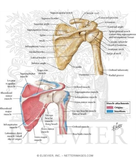
. Anatomical terms of bone edit on Wikidata In anatomy the scapula plural scapulae or scapulas also known as the shoulder bone shoulder blade wing bone speal bone or blade. It assists in forming the supraspinatous fossa and gives origin to part of the supraspinatus. Other structures labeled on the model include the ventricle 4 right atrium 2 left atrium 3 and the right and left truncus arteriosus 5.
The lower picture is a posterior from the rear view of the thorax scapula shown in red Details. As you can see there are also have a spine of scapula deltoid triceps brachii latissimus dorsi. We are pleased to provide you.
The ulna is on the medial side of the forearm and forms a hinge joint with the humerus at the elbow. Spread the knowledge. An MRI was performed on a healthy subject with a spin-echo T1 weighted acquisition.
This picture also contains humerus olecranon process of ulna deep to tendon and so on. Its inferior surface forms part of the infraspinatous fossa gives origin to a portion of the. Ventral rami of C5 to T1 course.
Tlso triplanar control rigid posterior frame and flexible soft anterior apron with straps closures and padding extends from sacrococcygeal junction to scapula lateral strength provided by pelvic thoracic and lateral frame pieces rotational strength provided by subclavicular extensions restricts gross trunk motion in sagittal coronal and transverse planes provides. The shoulder girdle includes three bonesthe scapula clavicle and humerus. Frog brain model dorsal view Lab-10 15.
MRI of the upper limb. Anatomical structures and specific regions are visible as dynamic labeled images. Malignant neoplasm of scapula and long bones of right upper limb C4002 Malignant neoplasm of scapula and long bones of left upper limb C4011 Malignant neoplasm of short bones of right upper limb C4012.
Root of spine shown in red. Emerges between anterior and middle scalenes courses through the posterior triangle of neck posterior to the clavicle before becoming closely associated with the axillary artery in the axilla before giving up its terminal branches or alternatively the roots emerge from behind the anterior scalenes to form 3 trunks. The humerus is the bone of the upper arm.
Anatomy note Youtube Channel Please Subscribe to support Anatomy note Odysee Channel Please Subscribe to Support. A useful mnemonic to remember the order of development is CRITOL or CRITOE see video. The vertebrate brain is divided into three main regions some of which are further subdivided.
The muscles of the shoulder support and produce the movements of the shoulder girdleThey attach the appendicular skeleton of the upper limb to the axial skeleton of the trunk. Data and DICOM images archived on a PACS Picture Archiving and Communicating System were processed and exported as JPEG images. It forms the ball and socket joint of the shoulder with the scapula and forms the elbow joint with the lower arm bones.
Muscles of the shoulder. This is useful information as the specific location of. The radius and ulna are the two bones of the forearm.
Its superior surface is concave. Anatomy terms allow us to describe the body and body motions more precisely. It presents two surfaces and three borders.
This image shows some of the major structures visible on the dorsal surface of the frog brain. Instead of your doctor simply saying that the patient knee hurts he or she can say that the patients knee hurts anterolaterally. Four of them are found on the anterior aspect of the shoulder whereas the rest are located on the shoulders posterior aspect and in the back.
The radius allows the forearm.

The Pectoral Girdle Anatomy And Physiology I
Scapula Posterior View With Labels Appendicular Skeleton Visual Atlas Page 5 Human Anatomy And Physiology Shoulder Anatomy Scapula

Scapula Posterior View Stock Photo Download Image Now Istock

Pin By Alper Doler On Scrub Life Anatomy Bones Scapula Bone Shoulder Anatomy

A P1lab Practical 2 Chapter 16 Flashcards Quizlet
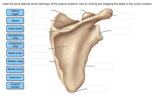
Solved Label The Bone Features Bone Markings Of The Chegg Com

The Pectoral Girdles Human Anatomy And Physiology Lab Bsb 141

Left Scapula Posterior View Labels 1 To 6 Indicate Areas Where Download Scientific Diagram
0 comments
Post a Comment